


Buchgreitz J, Buchgreitz M, Mortensen D, Bjørndal L
Guided access cavity preparation using cone-beam computed tomography and optical surface scans – an ex vivo study
International Endodontic Journal, 2016
For dentists and endodontists
Every endodontist has frozen in front of a calcified canal — and has watched another clinician solve the case they referred out.
Endoguide: guided endodontic access, calcified canals and digital planning.
10 lessons, 16 hours • 1 year of access • certificate • bonus e-book • no equipment purchase
English and Spanish subtitles
Start here: 2 minutes explain it all
The difference in practice
From conventional to guided access: more predictability and structure preserved.
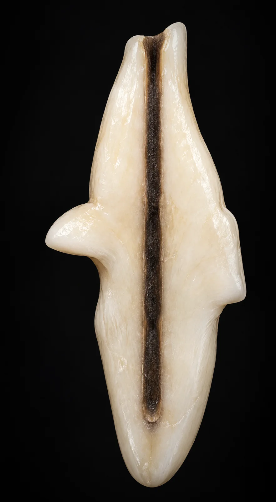
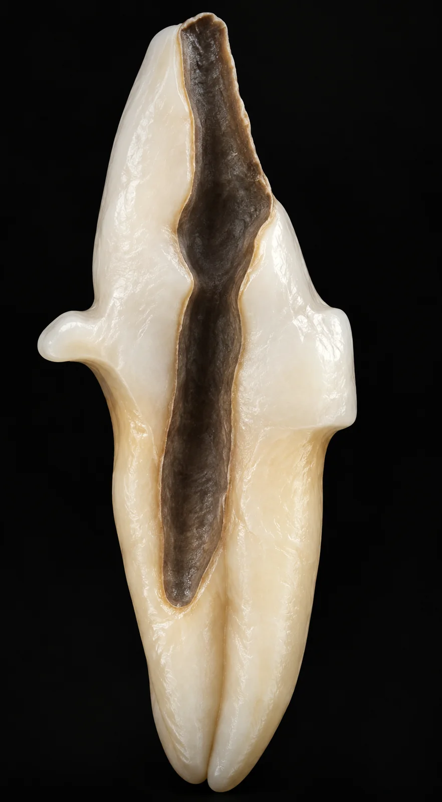
Schematic illustration. Access extent depends on each case anatomy.
The starting point
You know the case and you know the tooth can be saved. What you do not know is whether you get there without compromising the root.
Guided Endodontics moves that decision to before the access begins.
Perhaps this is your case
You open the tooth expecting the canal, and it is not where it should be.
You watch colleagues solve the cases you refer out, with the same training you have.
You remove more dentine than you would like, without knowing how much is left.
You have a case in your schedule you keep postponing, and you know exactly which one.
You assume you need to buy a CBCT unit and a printer to start.
You refer the case out and the patient does not come back — sometimes taking the family along.
None of this is a lack of technique. It is a lack of information at the exact moment the decision has to be made.
What Guided Endodontics is
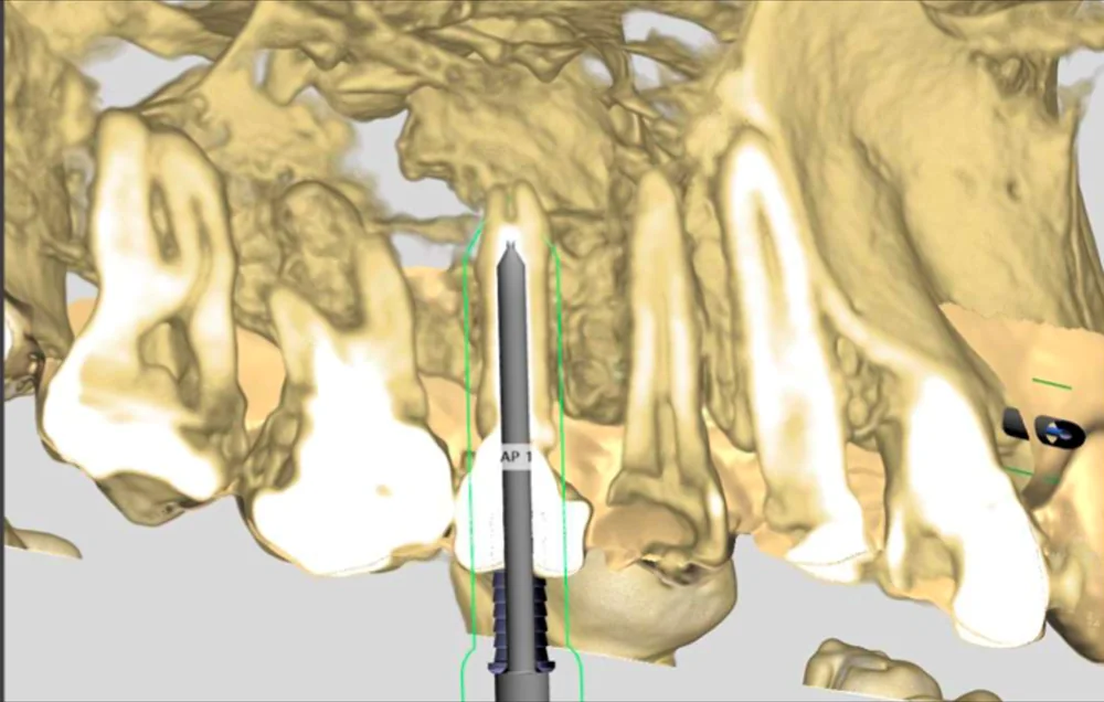
The trajectory is studied on the CBCT scan and carried to the chair by a guide.
The CBCT scan that reveals the actual anatomy of the case.
The reference from the teeth that will support the guide.
You define the trajectory before touching the tooth.
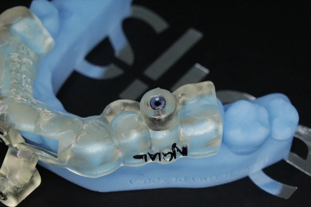
The plan becomes a physical object in your hand.
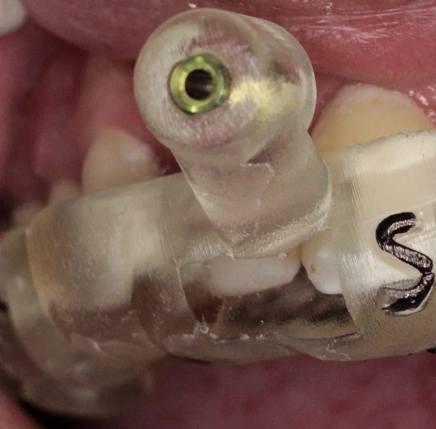
The access chairside, following what has already been decided.
From planning to chairside
You decide the trajectory on screen, unhurried, seeing the whole case. Then you seat the guide and carry out what has already been studied.
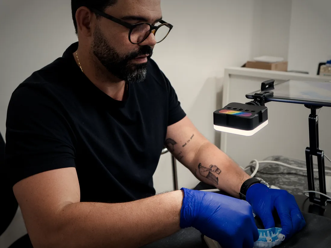
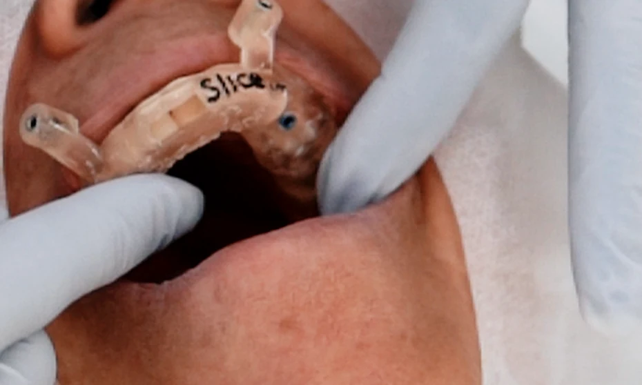
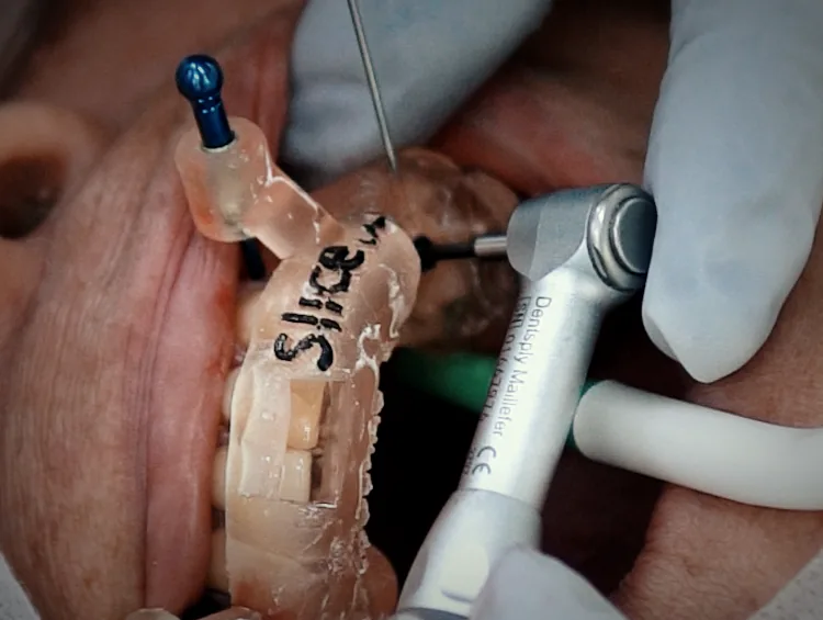
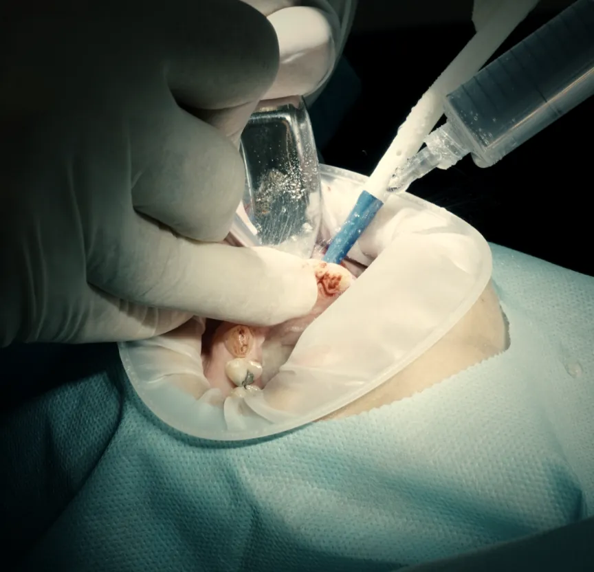
Actual laboratory stage and procedure, using Slice guides.
Curriculum
10 lessons and 16 hours, across five areas.
The principles and the digital workflow applied to Endodontics.
Indications, limitations and legal aspects.
From the imaging request to the trajectory defined on screen.
The guide at the chair, complications and guided microsurgery.
From planning to outcome.
Community, live sessions on YouTube and a channel to request one-to-one mentoring.
The starting point: how digital dentistry is organised and where Endodontics fits into that workflow.
How to request the right scan. This is the lesson that makes it possible to work with Endoguide without owning a CBCT unit: you learn the referral protocol for the radiology clinic.
Why the canal calcifies. Understanding the origin of the problem before deciding how to approach it.
Where the technique genuinely applies:
What the technique does not solve — and the legal dimension that accompanies the clinical decision. This is the lesson that separates those who indicate well from those who merely apply the tool.
The laboratory stage shown in practice — from planning to the finished guide.
Principles and techniques of microsurgery performed with the guided approach.
Cases performed and discussed, from planning to outcome.
Real cases
Three real cases, from the initial scan to follow-up. Tap to enlarge.
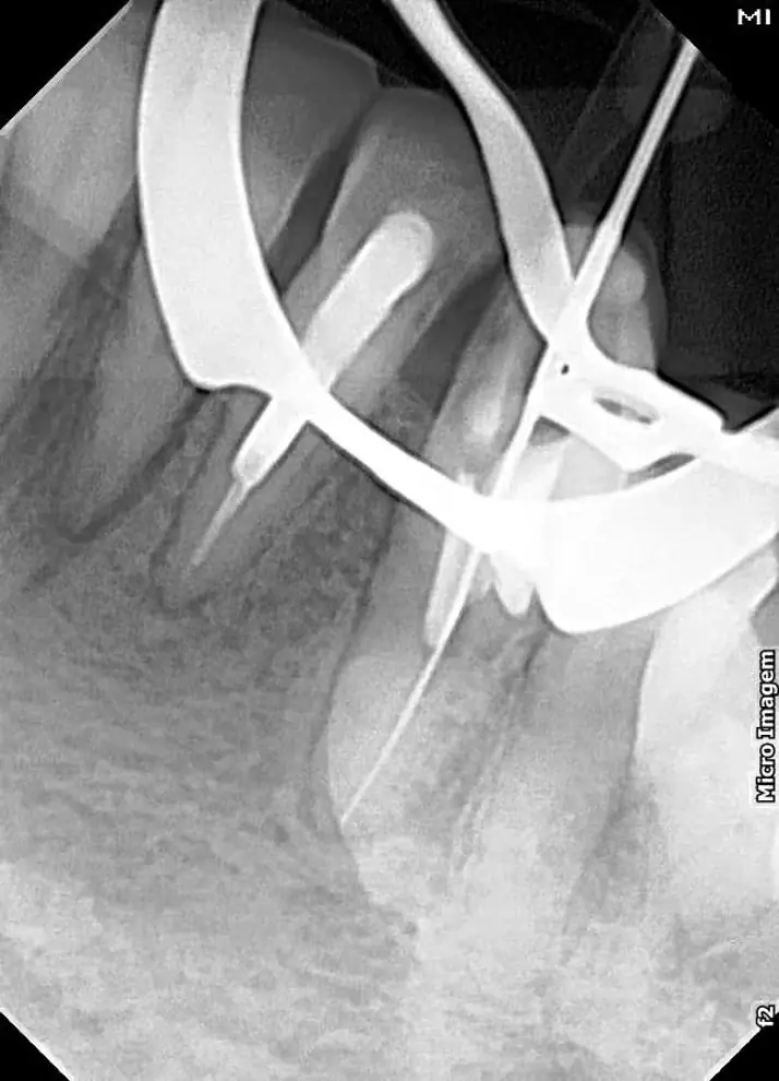
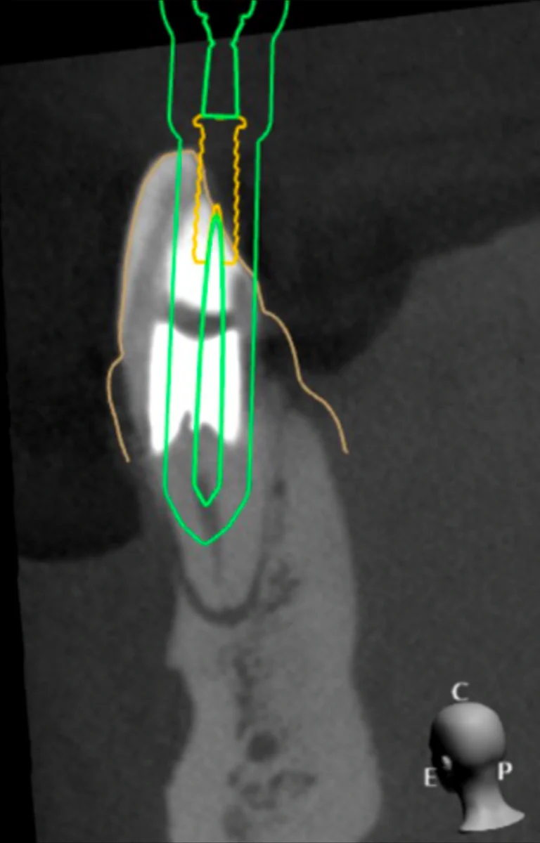
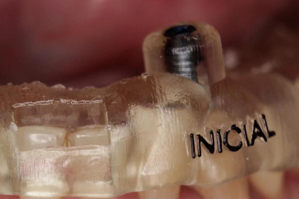
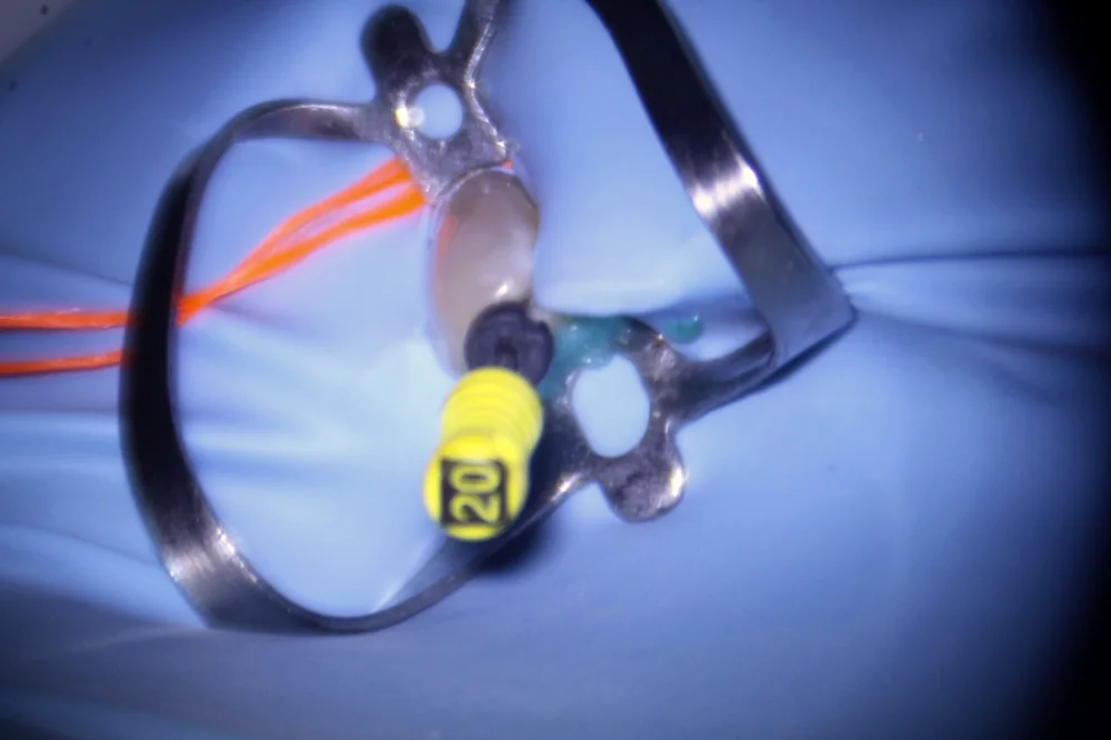
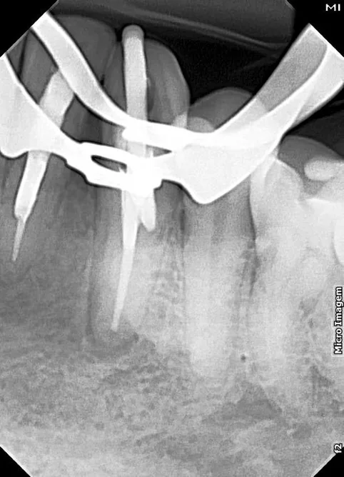
Swipe sideways to see all the stages.
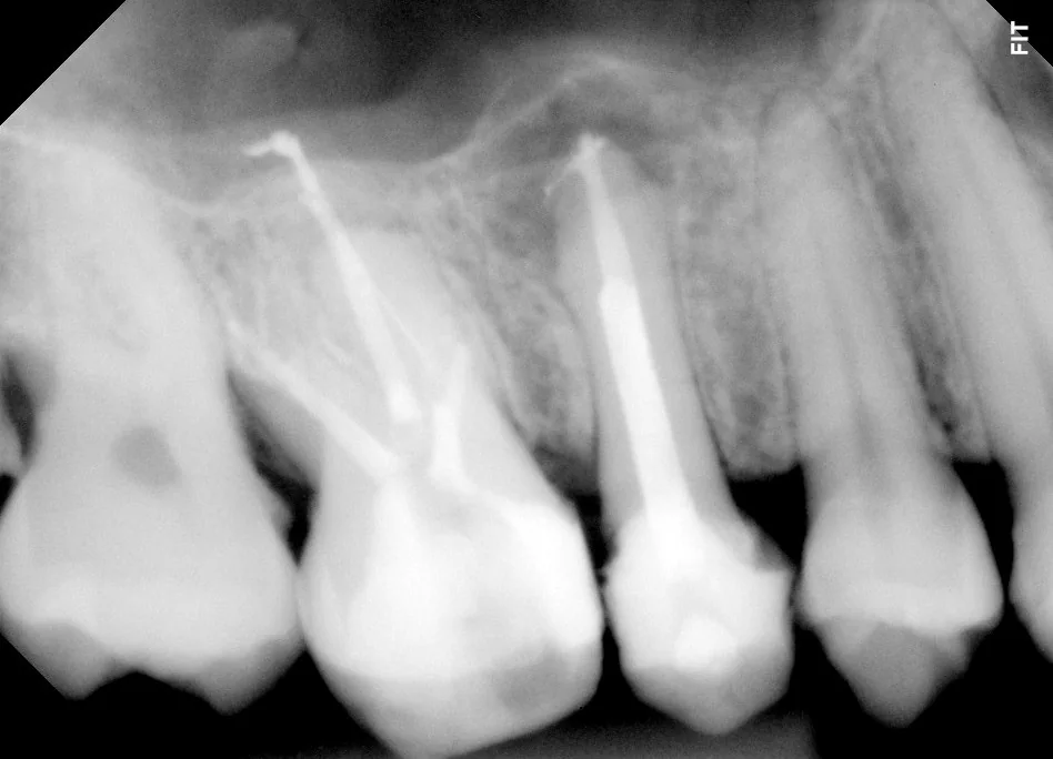
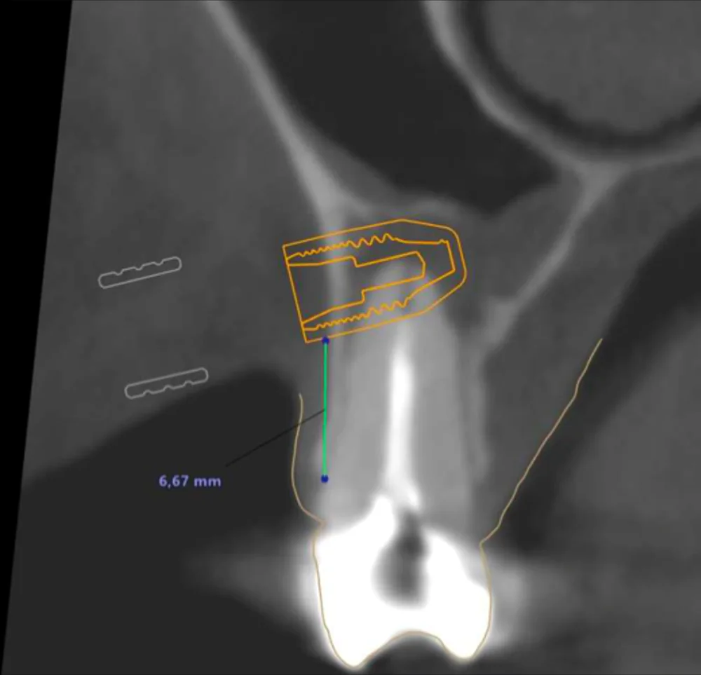
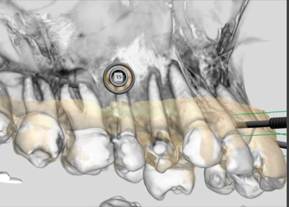
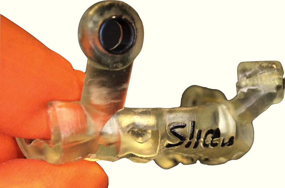
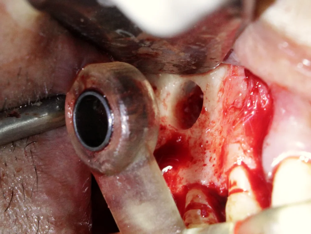
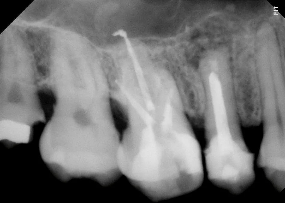
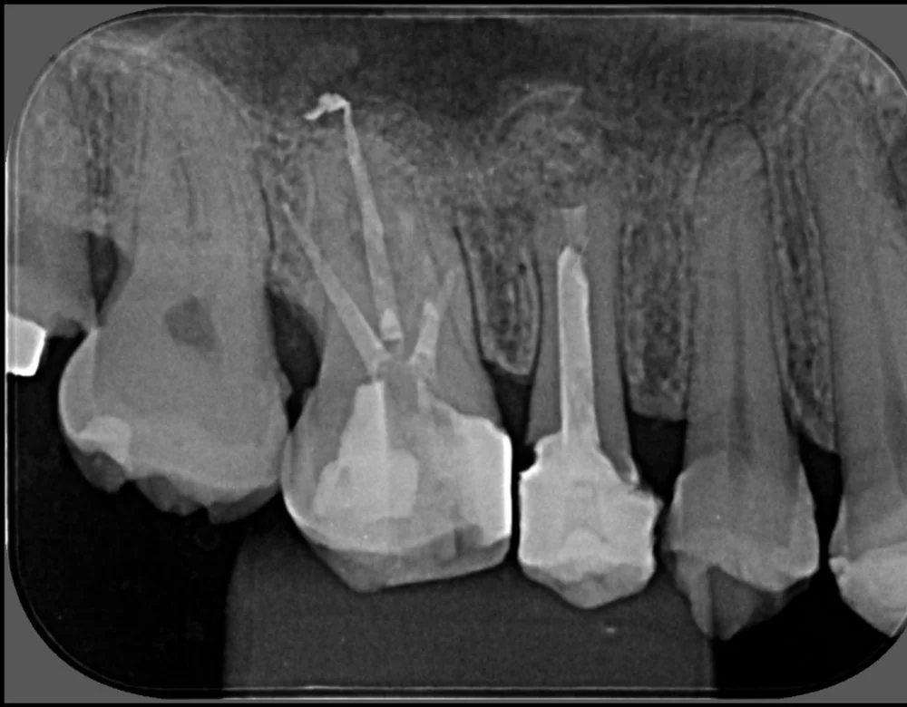
Swipe sideways to see all the stages.
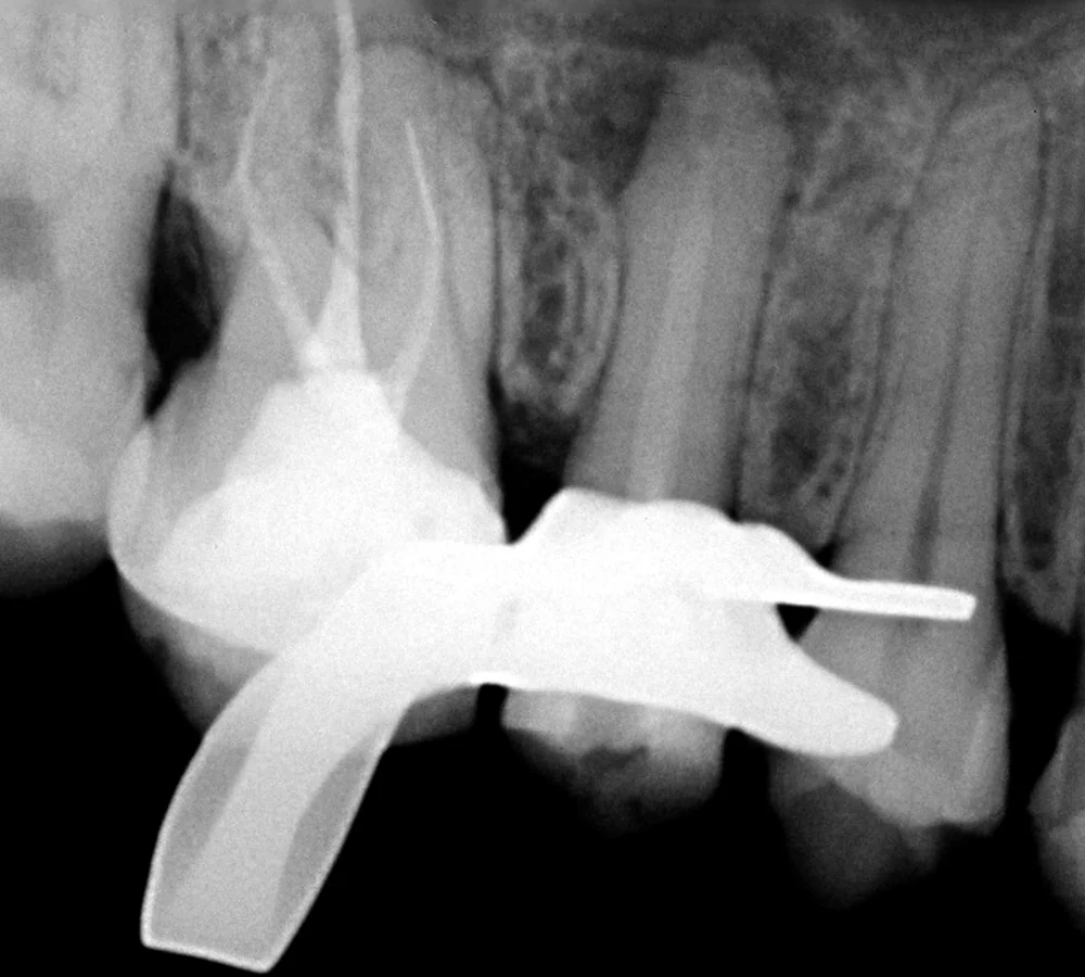
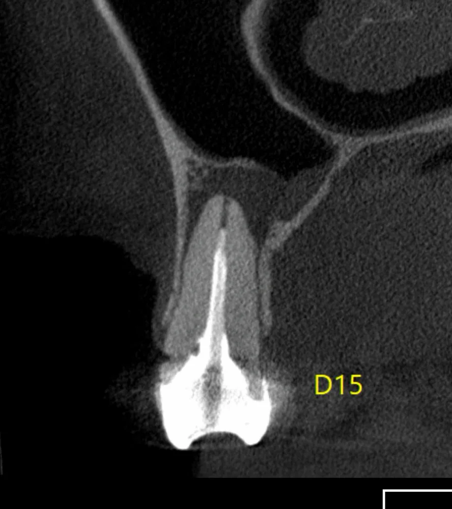
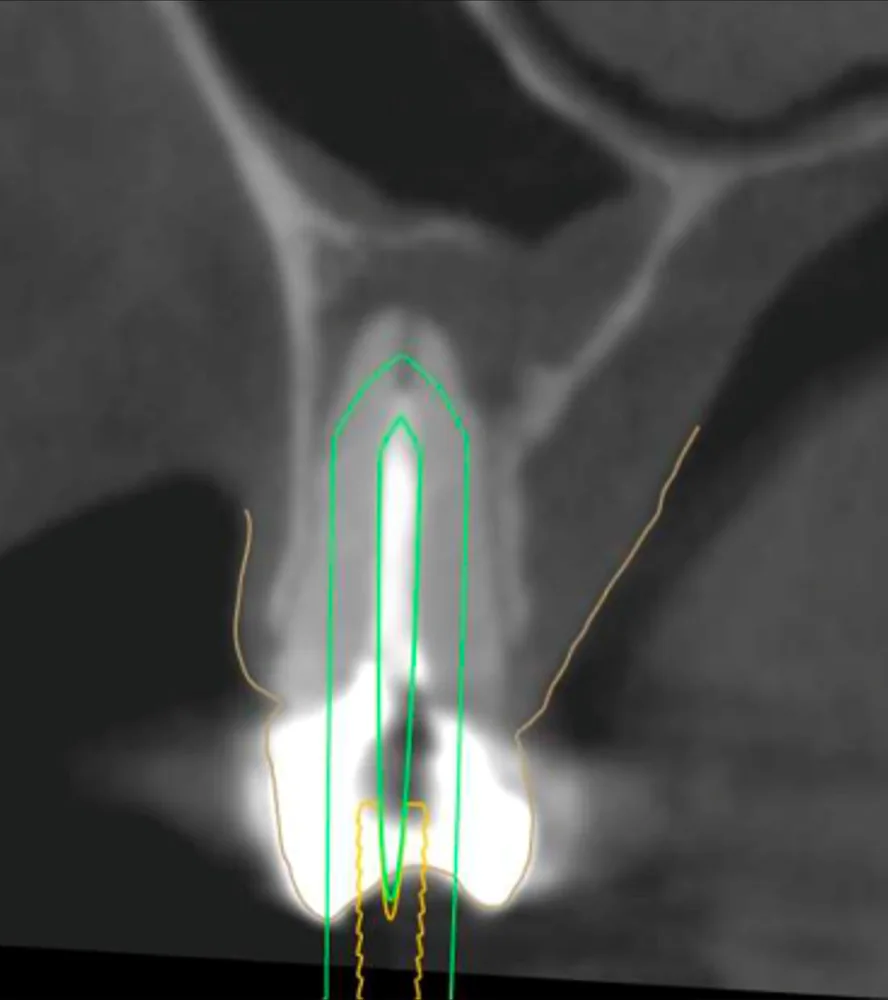
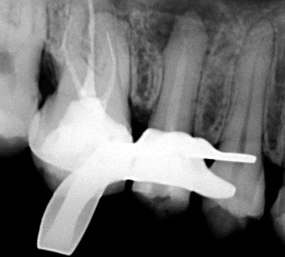
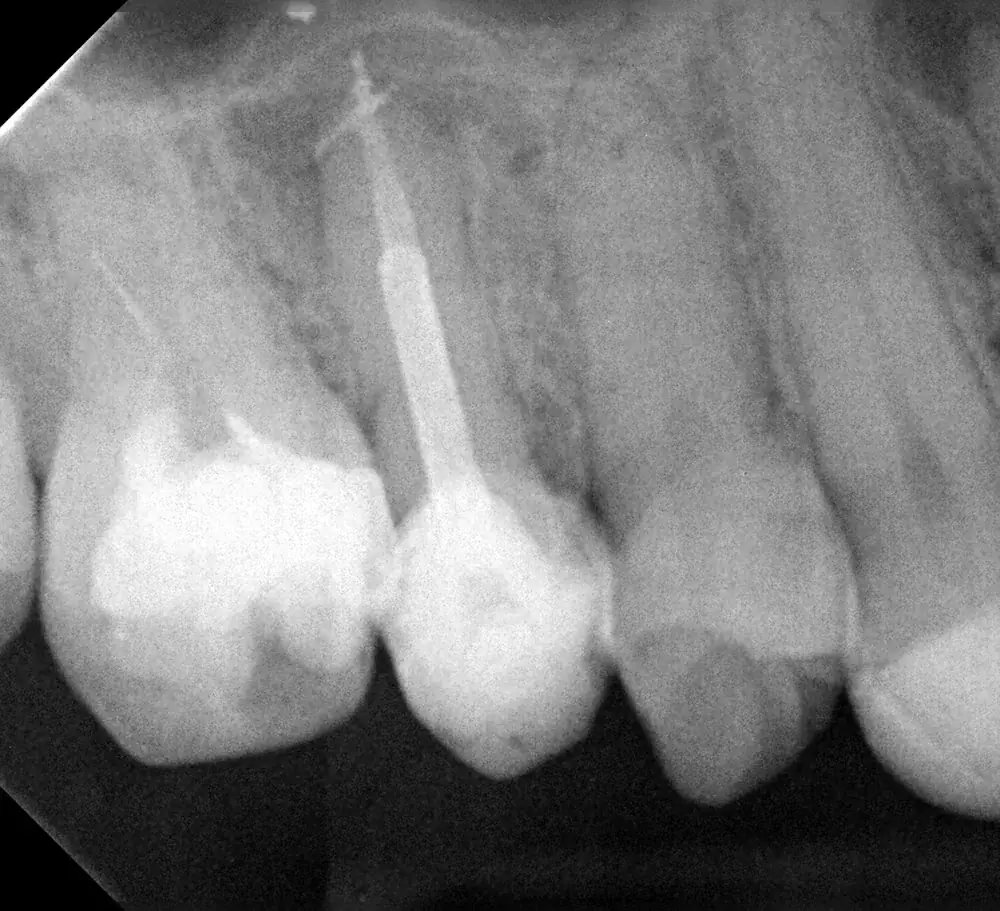
Swipe sideways to see all the stages.
Cases performed by Prof. Luiz Felipe Moreira, MSc.
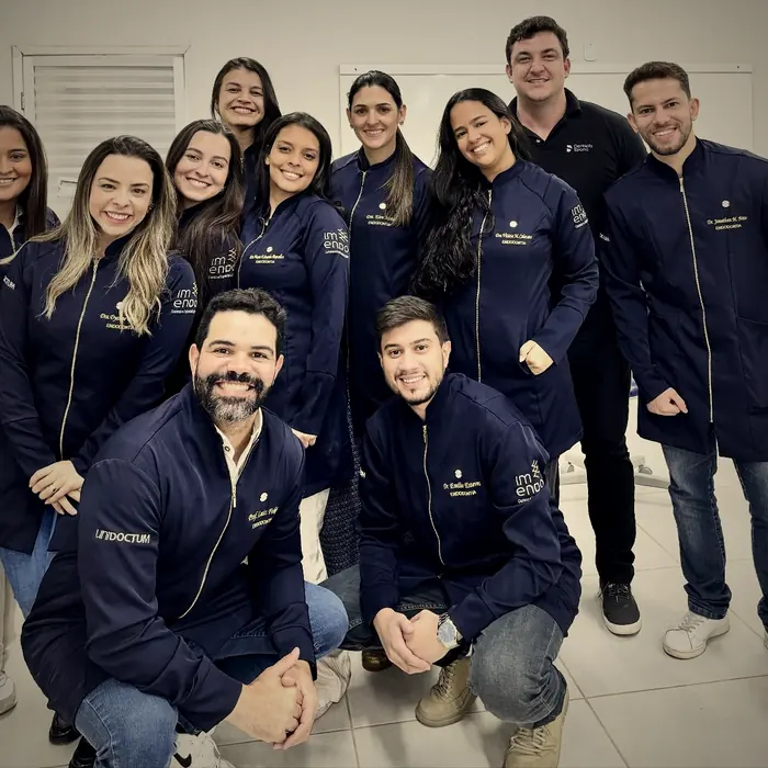
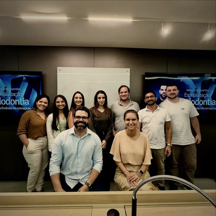
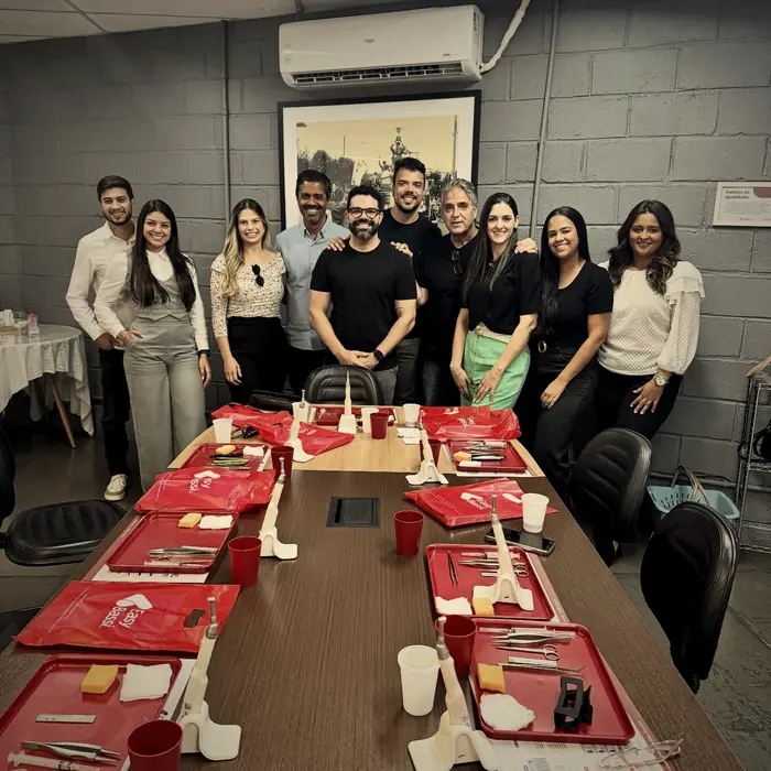
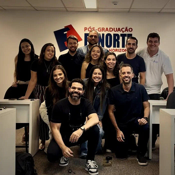
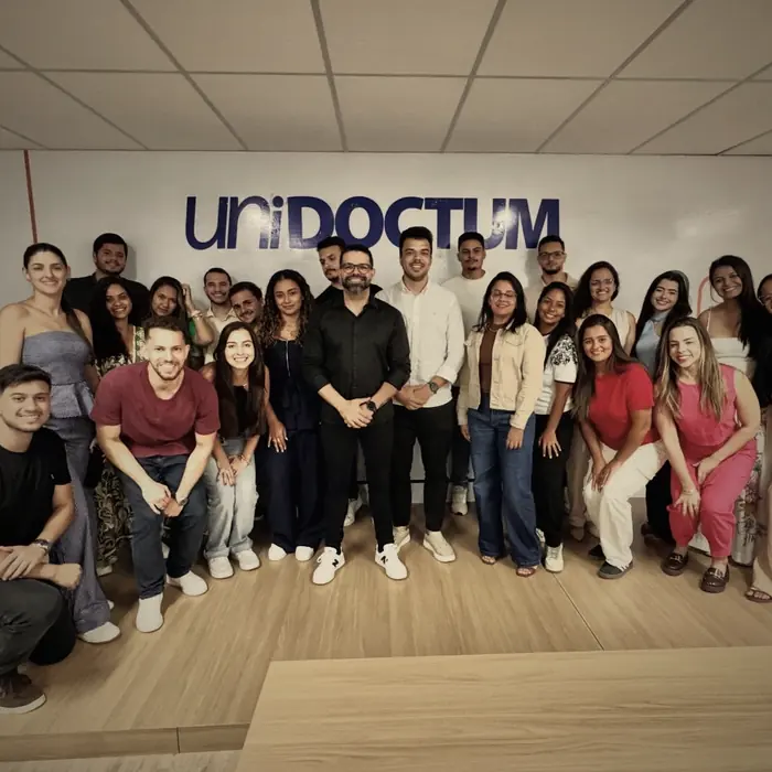
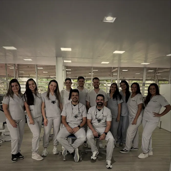
Proof from the classroom
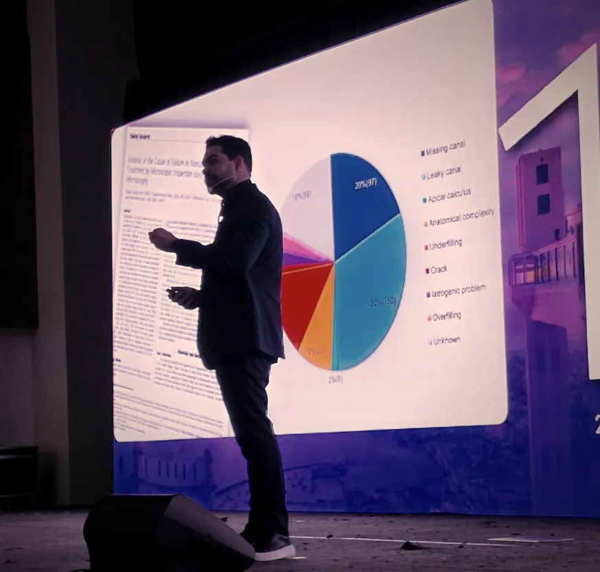
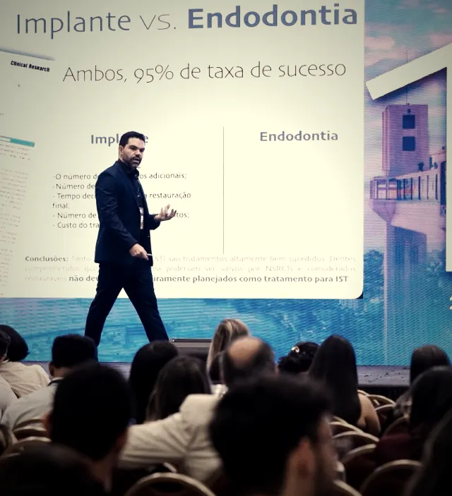
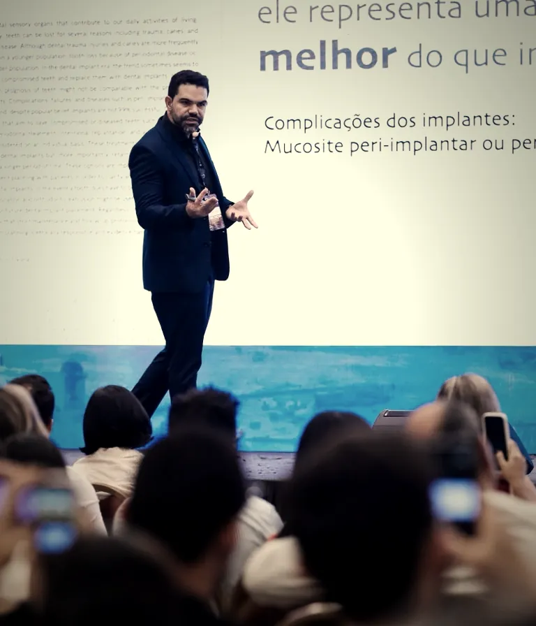
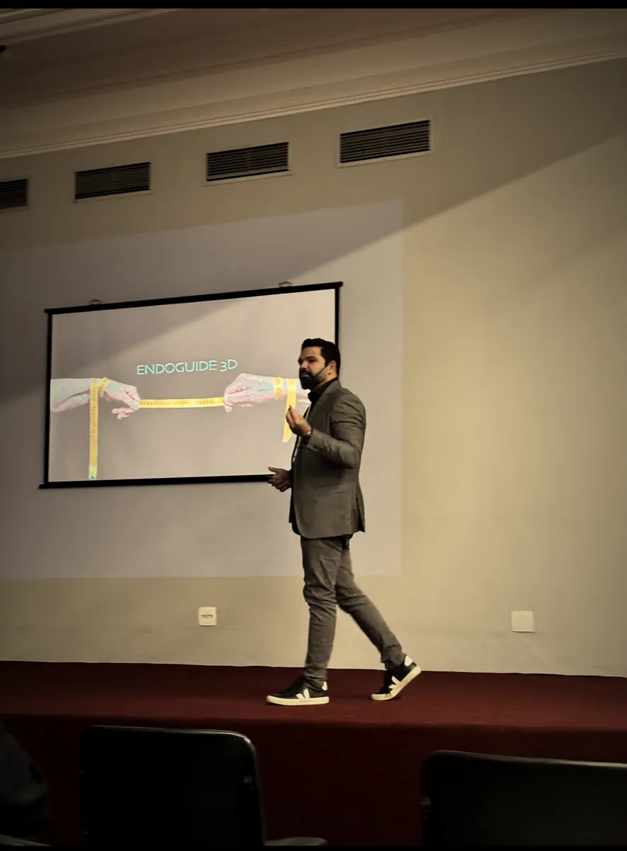
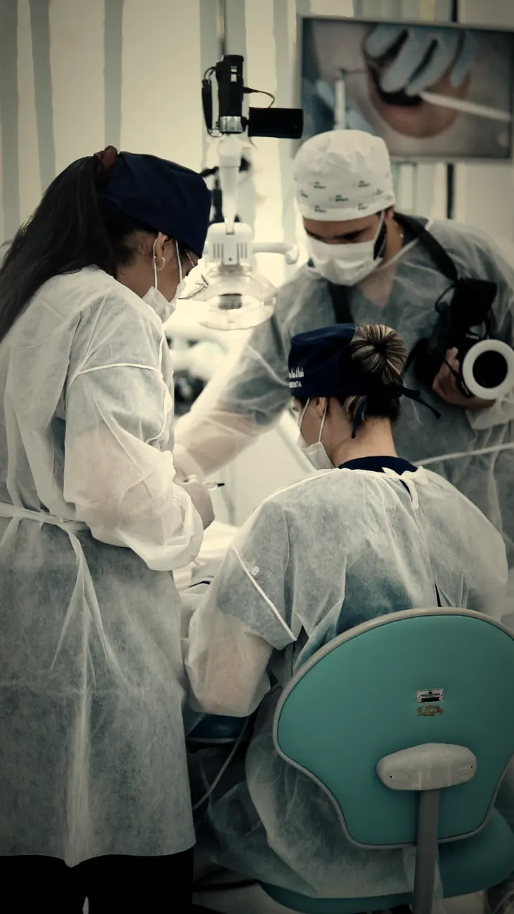
The course comes from clinical practice and from teaching the technique in person.
Swipe to see more
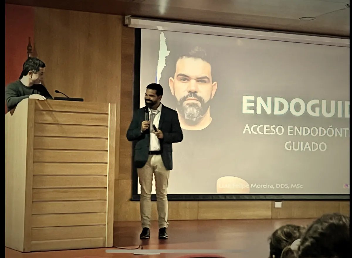
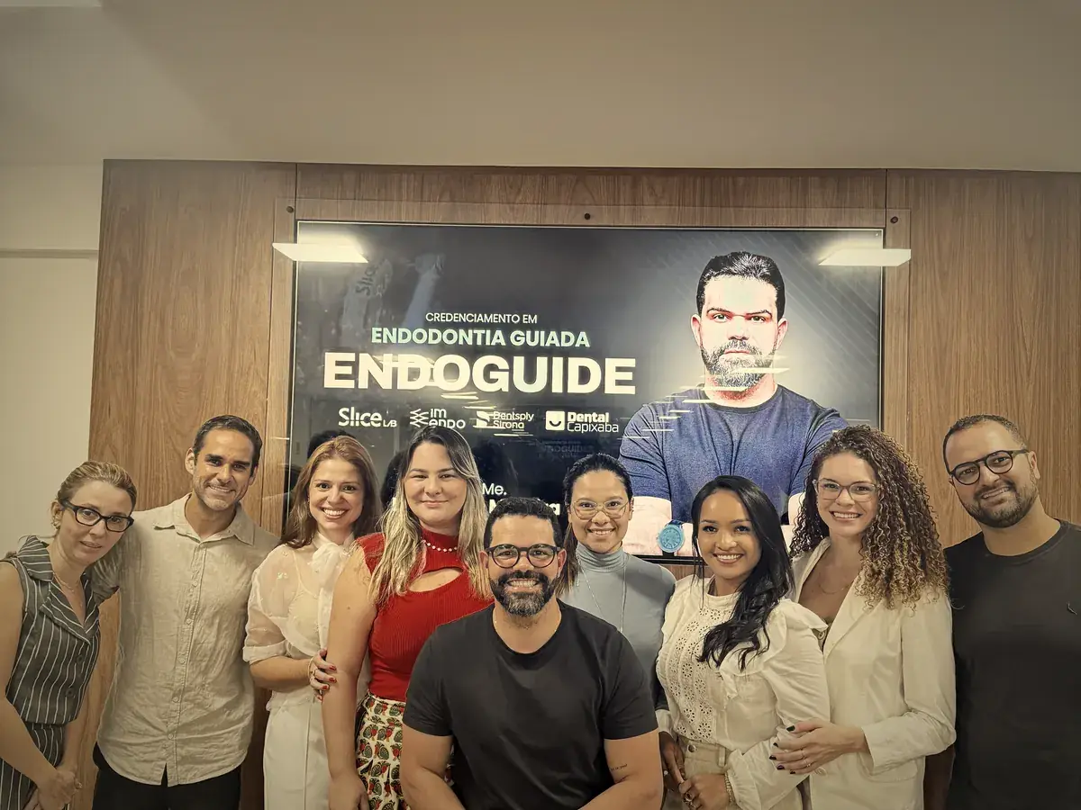
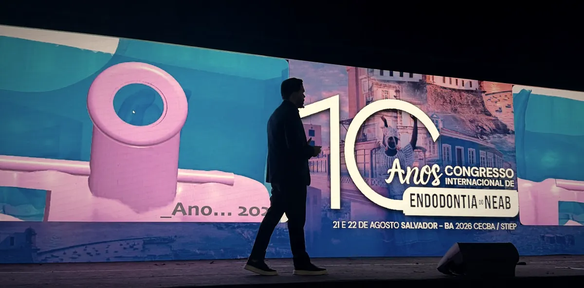
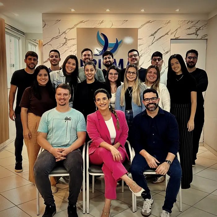
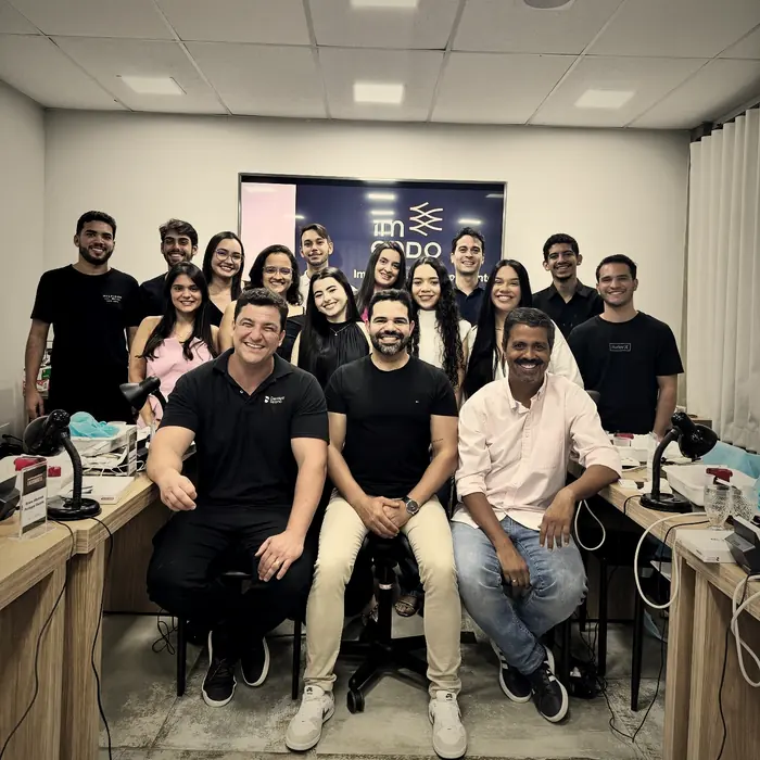
Classes trained

Brands and institutions along the way






Who will teach you
He has been doing Guided Endodontics since 2018 and teaches the technique to other clinicians. What you see in the lessons is what he uses in his own practice.
CRO-MG 39318
Scientific basis
23 studies published between 2016 and 2025. Nine by Brazilian authors.
Buchgreitz J, Buchgreitz M, Mortensen D, Bjørndal L
International Endodontic Journal, 2016
Zehnder MS, Connert T, Weiger R, Krastl G, Kühl S
International Endodontic Journal, 2016
van der Meer WJ, Vissink A, Ng YL, Gulabivala K
Journal of Dentistry, 2016
Connert T, Zehnder MS, Weiger R, Kühl S, Krastl G
Journal of Endodontics, 2017
Buchgreitz J, Buchgreitz M, Bjørndal L
International Endodontic Journal, 2019
Connert T, Krug R, Eggmann F, Emsermann I, ElAyouti A, Weiger R, Kühl S, Krastl G
Journal of Endodontics, 2019
Krastl G, Zehnder MS, Connert T, Weiger R, Kühl S
Dental Traumatology, 2016
Connert T, Zehnder MS, Amato M, Weiger R, Kühl S, Krastl G
International Endodontic Journal, 2017
Lara-Mendes STO, Barbosa CFM, Machado VC, Santa-Rosa CC
Journal of Endodontics, 2018
Lara-Mendes STO, Barbosa CFM, Santa-Rosa CC, Machado VC
Journal of Endodontics, 2018
Tavares WLF, Viana ACD, Machado VC, Henriques LCF, Ribeiro Sobrinho AP
Journal of Endodontics, 2018
Torres A, Shaheen E, Lambrechts P, Politis C, Jacobs R
International Endodontic Journal, 2018
Buchgreitz J, Buchgreitz M, Bjørndal L
Journal of Endodontics, 2019
Machado VC e cols.
Dental Press Endodontics, 2019
Maia LM, Machado VC, Silva NRFA, Brito Júnior M, Silveira RR, Moreira Júnior G, Ribeiro Sobrinho AP
Journal of Endodontics, 2019
Giacomino CM, Ray JJ, Wealleans JA
Journal of Endodontics, 2018
Maia LM, Moreira Júnior G, Albuquerque RC, Machado VC, Silva NRFA, Hauss DD, Silveira RR
The Journal of Prosthetic Dentistry, 2018
Casadei BA, Lara-Mendes STO, Barbosa CFM, Araújo CV, Freitas CA, Machado VC, Santa-Rosa CC
Australian Endodontic Journal, 2019
Chong BS, Dhesi M, Makdissi J
Quintessence International, 2019
Tavares WLF, Viana ACD, Ferreira MVL, Ferreira GC, Ferreira IC, Mesquita RA, Amaral RR
Journal of Endodontics, 2023
Moreira Junior G, Maia LM, Barbosa CFM, Toubes KMPS, Machado VC, Tavares WLF, Ribeiro Sobrinho AP
Iranian Endodontic Journal, 2025
Samiei M, Sabanik P, Tavakkoli Avval S
Iranian Endodontic Journal, 2025
Anderson J, Wealleans J, Ray J
International Endodontic Journal, 2018
Who it is for
From the fundamentals to the reasoning behind indication. It works for those starting out and for those who already treat these cases.
This training does not replace clinical training in Endodontics. It is aimed at professionals and students who wish to understand and incorporate the Guided Endodontics workflow within the limits of their own training and professional licensing.
Nor is it a shortcut: the technique requires study, appropriate case selection and a learning curve like any other.
What changes in your routine
No course guarantees a clinical outcome. What changes is the information you have when deciding.
The objection that stops most people
Imaging, planning and the guide stay with partners — just as you already do with radiographs.
What changes in your practice is the reasoning, not the assets.
Imaging, planning and guide costs exist, case by case. What does not exist is an upfront investment in equipment.
A phone. The lessons are recorded and stay on the platform, so you can watch on the device you already have, between appointments. No powerful computer or additional equipment is needed.
No. An intraoral scanner is not required to work with Endoguide.
No. The CBCT unit does not have to be yours — the imaging stage can be carried out at a radiology clinic, as already happens routinely in most practices.
You do not need to buy equipment to apply the technique. Guide fabrication is one of the stages that can be handled by partners, without you having to build the setup inside your practice.
Learning does not assume advanced CAD/CAM experience. The software is presented as a tool within a broader line of reasoning — not as the aim of the course.
Technology without clinical reasoning is just technology.
The course presents the planning workflow and the tools used at each stage.
You learn the workflow. Imaging, planning and the guide stay with partners.
I want the course →10 lessons • 16 hours • 1 year of access • 7-day guarantee
The offer
From your phone, between appointments. For 10 instalments of R$ 30.00, with a 7-day guarantee.
Online course
Guided Endodontics: from Planning to Execution
From R$ 997.00 to
or R$ 299.90 in full
Save R$ 697.00Secure payment • 7-day guarantee
Still have a question before deciding?
Chat on WhatsAppAll 10 lessons come with English subtitles. The bonus Endoguide e-book is fully translated — every chapter, table and reference. The instructor teaches in Portuguese, with subtitles throughout.
The course is sold in Brazil, so prices are listed in Brazilian reais. You will be charged in your own currency — the checkout converts the amount automatically at the exchange rate applied by your card issuer or payment provider on the day of purchase.
The final amount in your currency may vary slightly with the exchange rate and any international transaction fee charged by your bank.
Guarantee
If within 7 days you find it is not for you, the refund is requested on the platform itself. No justification, no conversation.
FAQ
Nearly fifty answers, grouped by topic. Tap the topic of your question.
Yes, always. Both the CBCT scan and the surface scan must cover the full arch, not only the region of the tooth — the guide rests on the adjacent teeth and needs them fully captured. In addition, the mouth recorded in the scan must be the same as on the day of the procedure: crowns, temporary restorations or fillings that will be removed should be taken out before scanning.
Endoguide is the application of Guided Endodontics in which a digitally produced guide directs access to the root canal system, based on virtual planning performed on three-dimensional imaging. The trajectory, angulation and depth are defined in software before the procedure.
Guided Endodontics is the approach that combines cone beam computed tomography, intraoral scanning or surface digitisation, virtual planning and a digitally produced guide to direct endodontic access in selected cases. The workflow runs from the imaging study to the printed guide, and from the guide to chairside access.
It is a root canal that has undergone progressive deposition of mineralised tissue, narrowing or obliterating its lumen — a condition also known as pulp canal obliteration. It may result from trauma, ageing, extensive restorations or chronic inflammation, and it makes locating the canal through the usual landmarks difficult.
In conventional access, the trajectory is defined during the procedure, based on visible anatomy and operator experience. In guided access, it is defined beforehand, in a digital environment, and reproduced chairside by the guide. The difference lies in the stage at which the decision is made.
The literature describes application in calcified and obliterated canals, fibre post removal and guided endodontic microsurgery. These are selected cases in which defining the trajectory in advance adds predictability — not a replacement for conventional access in every situation.
Yes. Cone beam computed tomography provides the internal anatomy of the root and canal, and it is on that dataset that the trajectory is planned. The scan does not have to be taken in your practice: it can be performed at a radiology clinic, and the course includes a lesson dedicated to the referral protocol.
Planning requires a surface reference on which the guide will rest. This may come from intraoral scanning or from other forms of digitisation. Owning a scanner is not necessary to work with the technique.
Yes. There has been literature published since 2016 in journals such as the Journal of Endodontics and the International Endodontic Journal, including accuracy studies, comparisons of tooth structure loss, case series and a systematic review on retreatment. This page gathers 23 of those publications with authors, journal, year and DOI.
No. No technique eliminates the risks of an endodontic procedure. Guided access adds planning to a stage that previously depended solely on interpretation during the clinical act, but diagnosis, indication, case selection and execution remain the clinician's responsibility. The course in fact includes one lesson on limitations and another on managing complications.
The purpose of the course is educational: to present the reasoning, the digital workflow and the clinical application so that you understand and can carry out the process. As with any technique, there is a learning curve, and application depends on appropriate case selection and each clinician's own practice. No outcome is guaranteed.
No. All you need is a phone. The lessons are recorded and remain available on the platform.
Yes. The content starts with understanding the concept and the workflow, before dealing with the execution of your first guide.
The course is aimed at dentists who perform or intend to perform endodontic treatment. Specialisation is not a formal prerequisite, but a clinical grounding in Endodontics matters in order to get the most from the content.
Yes. Specialisation and postgraduate students are among the course audiences, precisely because they are building their repertoire.
No. The whole point is to organise the process into a sequence that makes sense for those who do not yet work with this workflow.
No. You do not need a CBCT unit to work with Endoguide — the imaging stage can be done at a radiology clinic.
No. Owning an intraoral scanner is not necessary.
You do not need to buy equipment to apply the technique. Guide fabrication is one of the stages that can be handled by partners.
No. To follow the course, a phone is enough.
Yes. That is precisely why a scanner, CBCT unit or printer is not required: the imaging and guide fabrication stages can be handled by partners.
In equipment, nothing. There is no need to buy a scanner, CBCT unit, 3D printer or powerful computer. The imaging and guide fabrication stages can be handled by partners, as many practices already do with imaging studies. The investment is knowledge.
No investment in equipment is required. The imaging, planning and guide fabrication stages can be handled by partners, in the same way that many practices already outsource imaging today.
Broadly, the digital workflow involves the CBCT data (DICOM) and the surface record (STL). The course has a specific lesson on protocols for imaging referrals to the radiology clinic.
Learning does not assume advanced CAD/CAM experience.
Not to get started. Planning is one of the stages that can be handled with specialised partners. The course shows the workflow so that you understand and follow that stage, even when it is not done by you.
The lessons walk through the digital planning workflow from start to finish, presenting the tools as each stage arises — from reading the CBCT to defining the trajectory and the laboratory fabrication of the guide.
There is an entire module on managing complications: identifying perforations, instrument fractures and access difficulties, imaging diagnosis, prevention strategies and repair protocols, including surgical approaches when required.
Yes, and it has its own module. Understanding where Guided Endodontics does not apply is part of indicating it correctly.
There are 10 lessons and 16 hours of original content.
Yes. Digital planning is one of the pillars of the content and runs through the modules on indication, limitations, complication management and guided microsurgery.
Yes, on two fronts: a lesson on the aetiology of calcification and another on the indications for Guided Endodontics, which include calcification, fibre post removal and microsurgery.
Yes. There is a specific module on guided endodontic microsurgery, including custom surgical guides, visualisation of complex anatomy and instrumentation designed for the procedure.
Yes. There is a lesson dedicated to discussing clinical cases, and the complication management lesson also presents real cases with the strategies adopted in each situation.
Yes. There is a lesson demonstrating the laboratory execution of Guided Endodontics, plus the step-by-step application technique for bioceramic sealers.
Yes. All 10 lessons have English and Spanish subtitles, and the bonus e-book is fully translated into both languages — every chapter, table and reference. The lessons are taught in Portuguese, with subtitles throughout the course.
Yes. The course includes the Endoguide e-book, a reference for reviewing the main concepts of the training.
Yes. The course is fully online, accessed through the learning platform.
Yes. You study your way and on any device — phone, tablet or computer.
You have 1 year of access to the course from the moment it is released to you.
Yes, as many times as you wish, during your year of access.
Once payment is confirmed, access is released automatically. By card, confirmation is usually immediate; by bank slip or instant transfer, it depends on clearing.
Yes. The course issues a certificate of completion.
During your year of access, the content available on the platform is at your disposal, including anything published in that period.
Yes. In addition to the Guided Endodontics Community, the course has live sessions on YouTube and a channel to request one-to-one mentoring.
Yes. The course includes the Guided Endodontics Community, a space for exchange among students who are applying the workflow.
The course lessons are recorded, and in addition there are live sessions broadcast on the instructor's YouTube channel.
Yes. The course has a module dedicated to requesting one-to-one mentoring, with direct guidance from the instructor.
From R$ 997.00 down to 10 instalments of R$ 30.00, or R$ 299.90 in full, with a 7-day guarantee.
Yes. The entire purchase is processed by Hotmart, with the payment methods available at checkout.
Yes. There is a 7-day guarantee. Within that period, the refund is requested through the platform itself.
Final step
Ten lessons so you stop deciding the trajectory with the bur in your hand.
I want the course → See what you will learn10 × R$ 30.00 or R$ 299.90 in full • 1 year of access • 7-day guarantee